It has been a long while since the latest post in Onelephantsandbacteria, but we wanted to complete the series of Strangers in the Animal tree. That may represent a new renaissance on the blog, or at least, another small step to keep it alive.
Animals are fascinating examples of life on Earth and their evolutionary tree is full of imaginative and unique adaptations. Some animal groups are incredibly uncommon, mysterious creatures known by a handful of species which can challenge our current understanding of animal groups and their evolutionary history. These are the Strangers in the animal tree.
In the first issue, we went through the Cycliophora, small animals which have only been found attached to the mouth of shrimps, and the Micrognathozoa, minute animals that have some of the most complex jaw structures on Earth.
In the second issue, we went through the Placozoa, two-layered animals with genes that they don’t use, and the Orthonectida, animals that go through bizarre life cycles, more similar to fungi than other animals.
We end this series with Phoronida, as well as some small honorable mentions.
Phoronida

The Phoronida only have three different genus: Actinotrocha (only based on the larva), Phoronis and Phoronopsis, the latter only differentiated by a small fold at the base of the lophophore, the feather/tentacle-like structure that phoronids (and other lophophorates: such as brachiopods and bryozoans) have. With over 20 species, though, and even some potential fossil species, the phoronids are the best-known of all strangers in the animal tree.
As in many other invertebrates, the phoronida are marine-dwelling animals with two completely different stages:

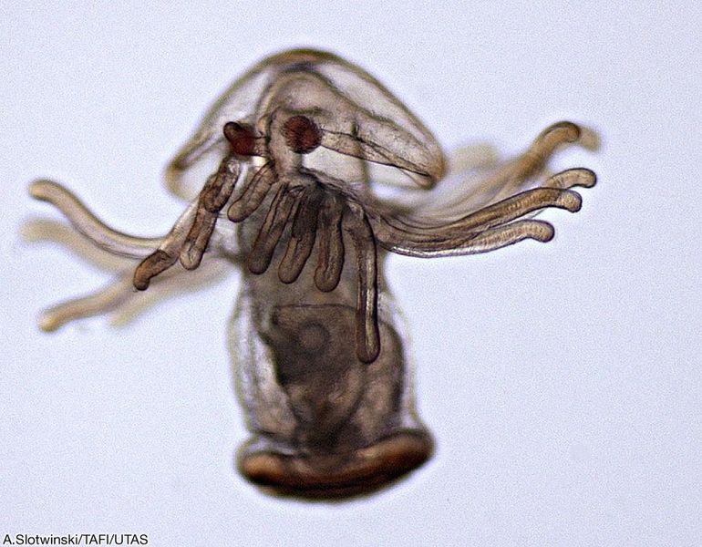
As larvae, phoronids are minute (less than 1 mm) inhabitants of the plankton, the vast communities of algae and microscopic animals that live close to the oceanic surface. These larvae look like small short tubes with their mouth covered by a hoot-like lobe and surrounded by a collar of tentacles. The rest of the body is short and simple, but ends with a series of small hairs (cilia), which they can adjust to change directions while swimming.
This stage can last from one to several months, until they start to perform an incredibly drastic metamorphosis that will lead them to their adult morphology.
Metamorphosis starts with a contraction of the body, creating a strong internal pressure. This pressure makes an internal lateral structure, the metasomal sac, to extend outside of the body. The metasomal sac will continue to grow and will eventually define the full body plan of the adult phoronid.
This lateral sac eventually becomes a tub-like structure that anchors the animal to the ground and creates a protective external layer of chitin. While the larva was free-swimming, the adult is now a sessile animal that filters particles from the water column. The old collar of tentacles in the actinotrocha larva is now the wide plumose structure known as the lophophore. The hoot, as well as the movable cilia, undergo cell death, and become reabsorbed. The adult stage is now mostly a ventral protrusion shaping the internal organs (such as the gut) into a “U” form, bringing the mouth and anus to the upper part of the tube, close to each other.
Where do Phoronids place in the animal tree? Phoronids are the closest group to brachiopods, a group of filter and deposit feeder animals that have two strong calcified valves, which look similar to bivalves to the untrained eye. Brachiopods are incredibly common in the fossil record, so much that they are used to infer past paleoclimates or even to set boundaries between eras in the geological timescale of the Earth. On the other hand, only the tube of phoronids is hard enough to allow some preservation and even then, it is difficult to tell if a fossil tube is really from a phoronid or any other tube-producing animal. There is a small list of fossil tubes which have been regarded as phoronids (e.g. Diorygma, Talpina) but none preserves soft tissues that would give information on their evolutionary history.
What happened to…
The Monoblastozoa ?

The Monoblastozoa are animals that may lie between the realm of zoology and cryptozoology (if they do not exist) or between zoology and protozoology (if they do exist).
Although described with authority, Manoblastozoa are only known from one description and a drawing from a single organism, Salinella salve. No specimen in a museum or collection exists of this animal.
Salinella was discovered in 1892 by the German biologist J. Frenzel, who found it in salt pans (areas where salty water has completely evaporated, leaving a wide layer of salt) in Argentina and since then, it has not been sighted again.
More contemporaneous attempts to find this enigmatic animal came from biologist Dr. Michael Schrödl in Munich some years ago, with no avail.
Was it all a prank? Frenzel was known to have written multiple monographs on microbiology and protozoology, where he described several species still valid today (Diplosiga, Mastigella, Phythelios…). It is difficult to imagine then, monoblastozoans being a prank, in a similar way to the Snouters from Steiner. In fact, most possibly Frenzel did not even think about the potential evolutionary importance of the monobalstozoans. Let me explain:

The main ground pattern of animals consists of a mouth a gut and anus (or a mouth-anus). All animals, (maybe with the exception of sponges, and the placozoans) have this pattern, which starts early in development, during gastrulation. In that early stages, the animal is a sphere of cells that bends inwards, creating the mouth or the anus and the gut. Therefore, if we were to stay inside the gut, in order to leave the animal, we would have to go through, at least, two layers of cells. In Salinella, though, there is only one layer of cells. This is suspicious, and may either indicate a very unique morphology, as in placozoans or, an early phase of multicellularity, aka, one of the first of animals.
The implications of this fact could be huge to our understanding of animal evolution. So far, nothing else is known from manoblastozoans, which remain a mystery.
Dendrogramma
When Dendrogramma was discovered in 2014 in a museum drawer in Australia, its bizarre shape made it unclassifiable. Its body was mushroom shaped and only a unique labyrinthic gut was visible. Many wrote about the possibilities of Dendrogramma being a new phylum altogether (including myself), a discovery that had not happened since the description in 1995 of the Cycliophora.

This striking pattern on the gut had hardly been seen in the animal kingdom either, and some speculatively related it to the spiral patterns found across Ediacaran animals. This was huge. Ediacaran animals are a group of animal-like (not all have been certified as animals) organisms that lived ca. 600 million years ago. Some resemble early cnidarians (jellyfishes) or poriferans (sponges), but others seem to have been an evolutionary end, a body plan that went extinct and never came. The consequences of Dendrogramma being a survivor of that Ediacaran fauna could have been enormous, indicating that Ediacaran animals survived and are still between us.

In 2016, an expedition to the southern Australian sea obtained new specimens (the previous had been found stored in formaldehyde, making them not suitable for molecular analysis) and looked at their genome. The answer was (drumroll): Dendrogramma is a cnidarian, a jellyfish. Although not the jellyfish one would normally picture, but a siphonophore.
A siphonophore is a type of colonial cnidarian (such as the Portuguese man o’war), where different members of the colony have specific functions, which translate into unique morphologies. Dendrogramma may be then, one of the members of an unknown cnidarian colony, more specifically a cormidia, a member specialized in making the whole colony float or protect it.
Dendrogramma most probably is not a descendant of the Ediacaran fauna (sad). But one mystery remains to be solved. If Dendrogramma is part of a colony, where is the colony? Known cormidia are a few millimetres, but Dendrogramma is almost two centimetres, which implies that a big magnificent colonial cnidarian is still hiding somewhere in the ocean.
Follow us on facebook and follow the author on Twitter for more!
References
Emig, C. C. (2010). Fossil Phoronida and their inferred ichnotaxa. Carnets de géologie.
Frenzel, J. (1892). Untersuchungen über die mikroskopische fauna Argentiniens.
Just, J., Kristensen, R. M., & Olesen, J. (2014). Dendrogramma, new genus, with two new non-bilaterian species from the marine bathyal of southeastern Australia (Animalia, Metazoa incertae sedis)–with similarities to some medusoids from the Precambrian Ediacara. PloS one, 9(9).
O’Hara, T. D., Hugall, A. F., MacIntosh, H., Naughton, K. M., Williams, A., & Moussalli, A. (2016). Dendrogramma is a siphonophore. Current Biology, 26(11), R457-R458.
Santagata, S. (2015). Phoronida. In Evolutionary developmental biology of invertebrates 2 (pp. 231-245). Springer, Vienna.
Temereva, E. N., & Malakhov, V. V. (2015). Metamorphic remodeling of morphology and the body cavity in Phoronopsis harmeri (Lophotrochozoa, Phoronida): the evolution of the phoronid body plan and life cycle. BMC evolutionary biology, 15(1), 229.
https://www.the-scientist.com/foundations/gone-missing-circa-1892-40412

2 Comments Add yours